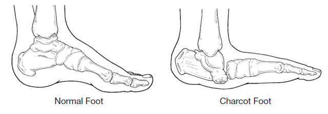
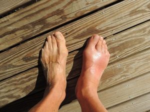
Charcot Foot Example

Charcot Foot

Charcot foot is a condition associated with neuropathy (nerve damage) to the foot.

It can lead to deformity, ulceration and even amputation. Although this problem is associated with many conditions that involve neuropathy to the feet, these include nerve damage caused by toxins (ethanol, drug related), infection (leprosy), as well as spinal cord damage and a number of other diseases (Parkinson’s disease, HIV, sarcoidosis, rheumatoid arthritis and psoriasis).

But by far the most common underlying problem is Diabetes, in over 20 years of looking at feet I have only seen one patient with a Charcot foot who was not diabetic.

There is still some, shall we say discussion in the medical profession as to the exact disease process, but most agree that weakened bones in the foot exacerbated by loss of feeling and sometimes trauma start off an inflammatory process. The current belief is that once the disease is triggered in a susceptible individual, there is uncontrolled inflammation in the foot. This inflammation leads to bone breaking down and is indirectly responsible for the progressive fracture and dislocation of joints in the foot.

Charcot foot symptoms, always include swelling of the foot often without a history of trauma. In the early stages there may be redness and warmth and sometimes pain. Although often diagnosed by examination and patient history x-ray is useful to confirm diagnosis. Changes to the bone that are seen on x-ray may be confused for a bone infection. A bone infection is very unlikely if the skin is intact and there is no ulcer present.

Treatment involves immobilising the foot usually with a cast or boot until the inflammation has stabilised. A Charcot episode usually results in changes to the structure of the foot often collapsing the arch of the foot resulting in a ‘rocker’ type foot. Following a Charcot episode an insole to support the foot and prevent further damage and charcot changes is required.

Contact me if you have any questions or would like further advice

Arch Pain

Arch Pain

This month I thought I would write about one of the most common causes of arch pain, Posterior Tibial tendinitis or Posterior Tibial dysfunction as it is sometimes known.

I see this condition a lot in my clinics and it can be a progressively debilitating condition. Which can, if left untreated result in rupture of the tendon.
 
It can present in several ways, a slow progressive onset with pain in the medial side of the arch starting from the middle of the arch. Sometimes the pain will progress around and behind the ankle and up the lower third of the leg. The condition is exacerbated by exercise and even walking can lead to discomfort. Or it can be sudden onset following particularly vigorous exercise or going down particularly hard on the affected foot.
 
Let’s just do a bit of anatomy before we come to the third presentation. Posterior tibial muscle is situated behind the shin bone (Tibia, as the name suggests) towards the outside of the leg. The tendon runs from the lower end of the muscle around the back of the inside ankle (Medial Malleolus) to attach to the Navicular a bone in the middle of the arch. This muscle is to a large extent responsible for pulling the arch of the foot up. So in those people with a low arch this muscle and tendon have to work a lot harder. If the foot flattens out (pronates) then with over use, the individual can experience pain at the attachment of the tendon (ensopathy) in the arch or as the tendon runs behind the ankle bone increased friction and irritation there as I have already mentioned can cause pain around the ankle and into the bottom of the leg.
 
The third presentation is pain on the top of the foot. But why? The attachment is under the arch? Well the body doesn’t like pain so if it hurts to use this tendon to maintain the integrity of the arch we then compensate and use the tendons on the top of the foot to try and do the same thing. Unfortunately they are not designed to do this and start to get inflamed themselves resulting in compensation pain on the top of the foot.
 
Finally there is rupture; this may not be dramatic and sudden as it can slowly progress from partial rupture with increasing lowering of the arch and spreading of the toes away from the midline of the body and turning out of the heel, to total rupture where the arch ends up touching the ground (weight bearing Navicular).
 
Occasionally rupture is sudden often taking the form of a sports injury the patient will feel it go and will require immediate surgical intervention within 24 hours before the tendon retracts up the leg.
 
So what to do? Well there is plenty. Rest and reduce the inflammation, be that with ice, anti-inflammatory drugs or as favoured in my office, Laser to the area. Then supporting the foot either with strapping or an orthotic (arch support)
 
In severe cases, an Air Cast which is a bit like a Ski Boot to immobilise the foot and give the tendon total rest. Usually Posterior Tibial tendinitis is diagnosed from symptoms and palpation, but where rupture or partial rupture is suspected MRI is very useful.
 
Philip Mann Podiatrist / Chiropodist 686 912 307 www.footpodiatrist.com